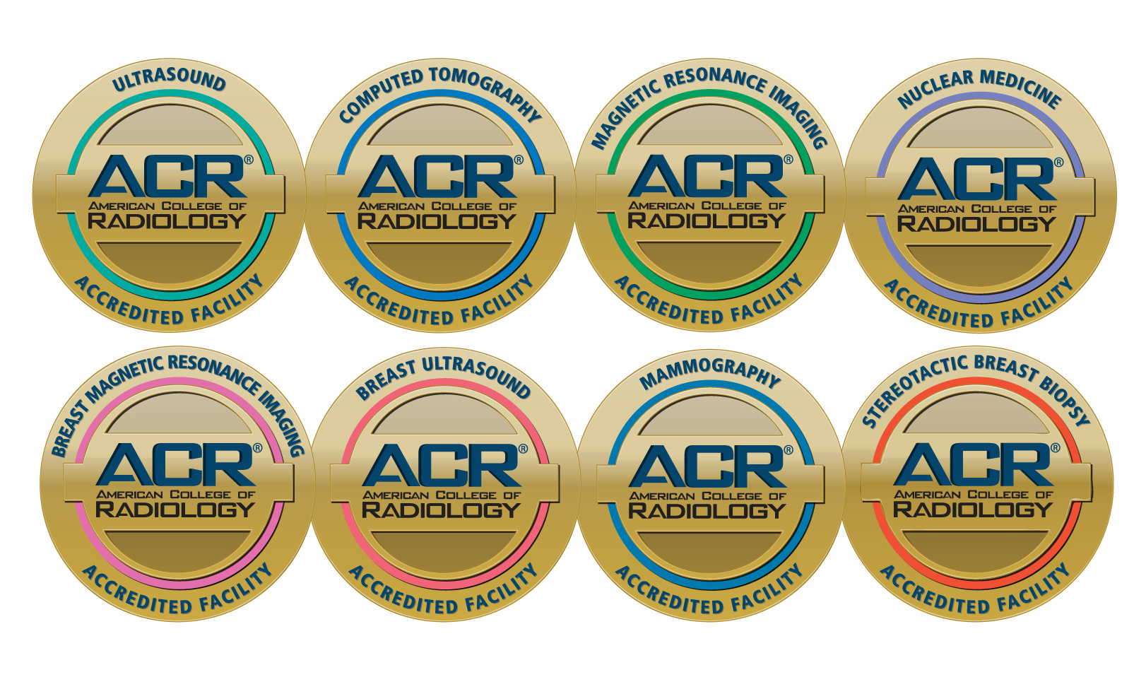
Our imaging department is fully digital, which means results are available to you and your doctor in less than 24 hours. Accreditation from the American College of Radiology (ACR) affirms we produce the highest quality of images with the lowest dosage of radiation.
Positron Emission Tomography (PET) is a state-of-the-art imaging tool that allows physicians to pinpoint the location of cancer within the body before making treatment recommendations. The highly sensitive PET scan images the biology of disorders at the molecular level, this works hand in hand with the CT scan which provides a detailed picture of the body’s internal anatomy.
Computed Tomography (CT/CAT Scan) uses special x-ray equipment to obtain image data from different angles around the body. It then uses computer processing of the information to show a cross-section of body tissues and organs. CT scans are the first line of defense for people experiencing the symptoms of a stroke to quickly determine if the patient can safely receive medication. Commonly used for diagnosing internal injuries, cancer, tumors, or kidney stones, CT is also a trauma modality.
Magnetic Resonance Imaging (MRI) uses radio waves and a strong magnetic field rather than x-rays to provide remarkably clear and detailed pictures of internal organs and tissues. The technique has proven very valuable for the diagnosis of a broad range of conditions in all parts of the body, including cancer, heart and vascular disease, stroke, and joint and musculoskeletal disorders.
Mammography is a specific type of imaging using a low-dose x-ray system for examination of the breasts. Pullman Regional Hospital offers the most advanced technology with digital mammography. Digital mammography is 25 percent more effective at detecting abnormalities in women with dense breasts than conventional mammography. While other diagnostic tests are sometimes necessary, the mammogram is the first line of defense against breast cancer.
Nuclear Medicine imaging might sound ominous, but it's very safe; it is also incredibly effective in giving medical professionals insight into how the body is functioning. Unlike traditional anatomical imaging, such as computed tomography (CT) or magnetic resonance imaging (MRI), nuclear medicine exams show how the body functions abnormally when a disease or dysfunction is present. The amount of radiation used in a nuclear medicine exam is very small, and there are no measureable side effects. At Pullman Regional Hospital, we use radiation with a short half-life, meaning there is even less exposure to the patient.
Ultrasound Imaging, also called ultrasound scanning or sonography, is a method of “seeing” inside the human body through the use of high-frequency sound waves. The sound waves are recorded and displayed as a real-time visual image. No radiation is involved in ultrasound imaging. Ultrasound is a useful way of examining many of the body’s internal organs, including the heart, liver, gallbladder, spleen, pancreas, kidneys, and bladder. Ultrasound, as most people know, is the preferred method used to visualize an unborn fetus.
X-Ray Imaging, or radiography, is the oldest and most frequently used form of medical imaging. For nearly a century, diagnostic images have been created by passing small, highly controlled amounts of radiation through the human body, capturing the resulting shadows and reflections on a "digital" plate. X-rays allow the physician to view and assess broken bones, a cracked skull or injured backbone. X-rays also play a key role in orthopedic surgery and the treatment of sports injuries.


